Dual-galvanometer scanning
Two galvano scanners steer the beam in XY or XYZ for full-field scans, targeted ROI scans, and park mode point measurements.

CoraScan is a compact fiber-coupled laser-scanning confocal scan head for configurable microscopy, three-dimensional surface profiling, fluorescence imaging, and OEM instrument development.
The current configuration is designed around the Nikon Eclipse Ti2-E inverted microscope, using 1x to 100x objectives. CoraScan keeps the optical scanning core compact while leaving the laser and detector chain open to the application.
Two galvano scanners steer the beam in XY or XYZ for full-field scans, targeted ROI scans, and park mode point measurements.
Route the descanned signal through the FC/APC output to an APD, PMT, or photon-counting setup selected for the experiment.
Select a 25, 50, 75, or 100 micrometer pinhole and specify filters with a broadband beam splitter or wavelength-selective dichroic.
Acquire full, ROI, and park measurements, including XY images and XYZ stacks, with the supplied control software.
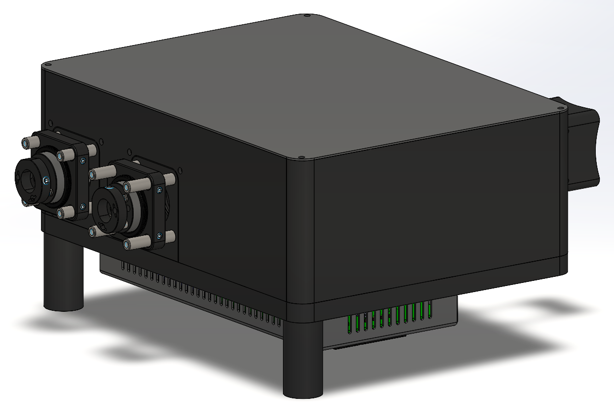
Configuration details below are based on the current CoraScan brochure. Laser source and detector are selected separately.
| Scanning | |
|---|---|
| Scanner | Dual-axis galvano scanner |
| Scanning modes | XY and XYZ |
| Scan patterns | Full scan, ROI scan, and park mode |
| Image format | Up to 2000 x 2000 pixels |
| Frame rate | 1.0 fps at 512 x 512 pixels, bidirectional |
| Pixel dwell time | 1 to 8 microseconds |
| Optics and ports | |
| Laser input | 1 x FC/APC fiber port |
| Signal output | 1 x FC/APC fiber port |
| Objective range | 1x to 100x microscope objectives |
| Confocal pinhole | 25, 50, 75, or 100 micrometers |
| Excitation and emission filters | Specified per application |
| Beam splitter or dichroic | Broadband beam splitter or wavelength-selective dichroic |
| Detection | External APD, PMT, or photon counter via signal fiber |
| Compatibility and software | |
| Microscope platform | Nikon Eclipse Ti2-E inverted microscope |
| Software | Ready-to-use acquisition and control software |
| Software functions | Full, ROI, and park acquisition with XY and XYZ stacks |
| Power supply | 24 V DC, 2.5 A |
Select the confocal pinhole to trade optical sectioning against signal throughput.
25 um50 um75 um100 umAdd excitation and emission filters matched to the laser line, sample, and detection band.
Choose a broadband beam splitter or wavelength-selective dichroic to suit the excitation and emission paths.
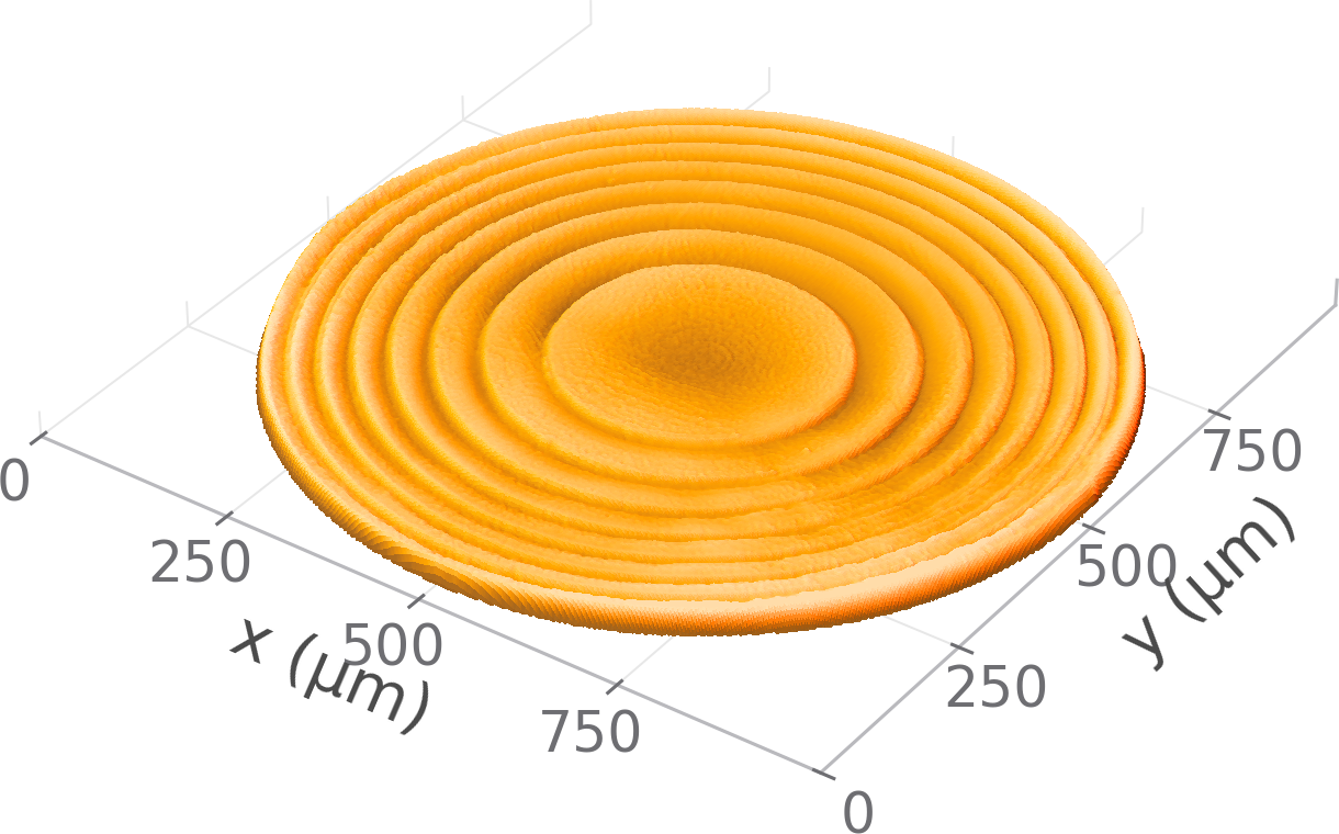
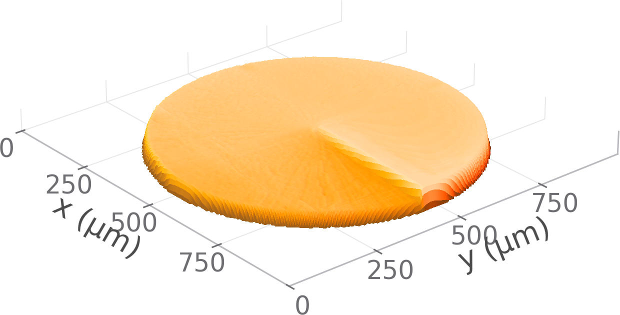
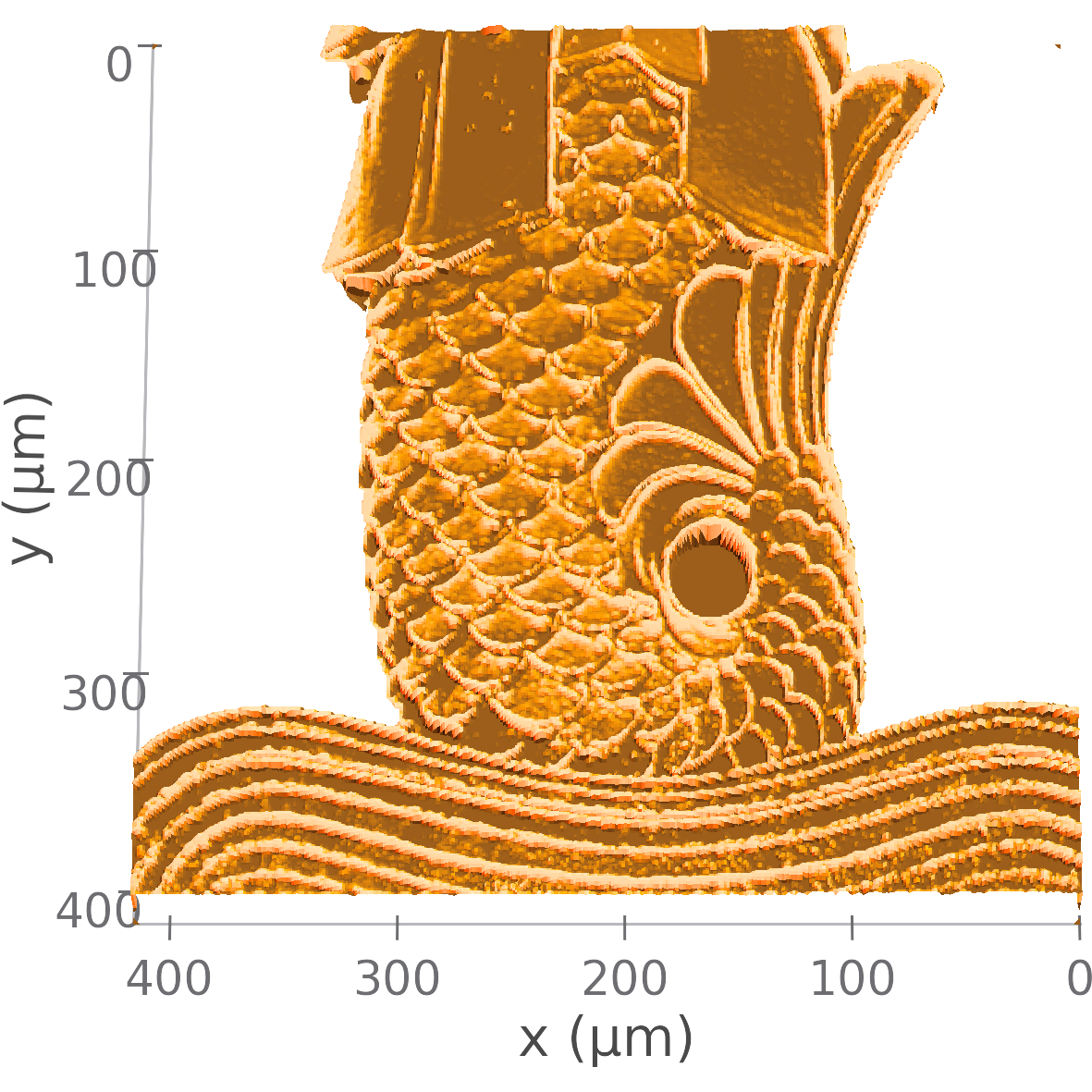
Demonstration results measured with the scan head on fabricated grayscale microstructures.
Share your microscope, excitation wavelength, detector, sample, field of view, and measurement objective. HX Technology will help define a suitable CoraScan configuration.